Eye
Man can identify its surroundings through the senses of the five, namely: hearing, smell, taste, and touch, and finally the sense of sight , and represents the eye that works like a digital camera in a lot of qualities, and these similarities as follows: [1]
- The light is focused primarily on the cornea of the eye, which is represented by the camera lens.
- The iris controls the amount of light that reaches the back of the eye. By automatically changing the size of the pupil , which represents the diaphragm in the camera.
- The focus of light is increased automatically on nearby objects through a process called accommodation eye. And this is done through the eye's crystal lens, which is located directly behind the pupil , which resembles an automatic focus lens inside the camera.
- The focused light by the cornea and the crystalline lens reaches the retina . Which is similar to the image sensor inside the camera, the retina works on converting light into electronic signals, and then transmits it to the optic nerve to the visual cortex.
Eye ingredients
The human eye resembles a sphere, with a slight indentation at the front, and the eye consists of three basic layers located on top of each other. Which in turn forms the eyeball, and the eye is divided from the inside into three main chambers, and what follows is an explanation of all these components: [2]
- The layers of the eye, and the basic layers of the eye can be summarized as follows:
- The sclera: is the white part of the eye, and it forms a wall to support the eyeball, and this layer increases at the area surrounding the optic nerve, and this layer is covered by the conjunctiva ; It is a mucous membrane that helps to moisturize the eye, and the sclera layer extends to connect with the cornea , [3] as for the cornea ; It represents the transparent layer of the sclera, and it works to convert images of things that a person sees into light waves that travel into the eye, and both the transparency of the cornea, its shape, and the smoothness of its surface are important in improving the function of the eye. [4]
- Chorionic: choroid layer is located between the retina and solid layers . This is a much thicker layer at the back of the eye; It is 0.2 mm, and its thickness begins to gradually decrease around the eye to 0.1 mm. This layer contains blood vessels in the eye, in addition to it contains the pigment epithelium of the retina of the eye, so the retina provides the oxygen and nutrients needed for the outer layer of the retina. This layer is on the melanin pigment that absorbs light and reduces its reflection inside the eye. Because of its negative impact on the process of vision, and this dye protects the blood vessels located in the layer of the placenta from the influence of negative light in it, and the placenta forms the uveal tract. Which in turn includes all of the irisAnd the ciliary body , and they can be detailed as follows: [5]
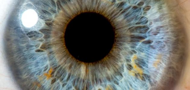
- The iris: , is the colored part of the eye, which regulates the amount of light waves passing into the eye, and the iris contains in the middle of it an opening known as the pupil, which passes the light organized by the iris. [6]
- Ciliary body: ciliary bodies stretch or contract; This helps the lens of the eye change shape and obtain a more accurate focus. [6]
- The retina: It is the third and last layer of the layers of the eye, and it is worth noting that the retina contains millions of light-sensitive cells, and these cells form in two main shapes: conical or rods. Rods sense monochromatic colors during dim light. It is concentrated within a region called the central fossa . Which helps to see in a high and sharp focus, when light comes into contact with these sensitive cells, they are converted into electrical signals that are transmitted to the brain by the optic nerve . [7]
- Eye Compartments : The rooms are divided into three sections, namely: [2]
- The anterior chamber of eyeball, which is the front part of the eye, and is located between the cornea and the iris.
- The posterior chamber of eyeball, located between the iris and the lens.
- The vitreous chamber, located between the lens and the back of the eye.
- The protective components of the eye: There are several other parts of the eye whose function is to protect the eye, including: [8]
- The bony cavity of the eye: the eye is protected by the presence of a cavity that allows it to move freely despite the presence of blood vessels, nerves, and muscles in the eye.
- Eyelashes: They are short, strong hair that grows on the edges of the eyelid, and they act as a barrier to the eye. It prevents insects and foreign bodies from entering the eye.
- Eyelids: They are thin sheets of skin and muscles located above and below the eye, and protect the eye from insects and foreign bodies, or when intense light shines on the eye, as they close automatically and very quickly for the purpose of protection.
- The conjunctiva: It is located on the back surface of the eyelid and covers the front surface of the eye to the edge of the cornea, and it works to protect the tissues beneath it.
- Tears: they are salt water that constantly floods the surface of the eye to maintain its moisture, and it conveys oxygen and food to the cornea, and tears contain antibodies. Which reduces the incidence of infection.
As a person ages, he is more likely to develop eye diseases associated with aging, and among these diseases are the following: [9]
- Macular degeneration: It is a disease associated with aging, and it leads to poor focus of vision for things, and during certain tasks such as reading and driving a car.
- Cataract: , also called cataract , it means a darkening of the lens of the eye; This causes a flare in vision, and thus blurred vision.
- Eye Diabetes: , and is one of the complications of the disease of diabetes , and the main causes blinding, occurs when blood vessels in the retina of the eye damage due to diabetes.
- Water Blue: , a group of diseases associated with higher eye pressure may damage the optic nerve; This affects side vision, and may cause vision loss.
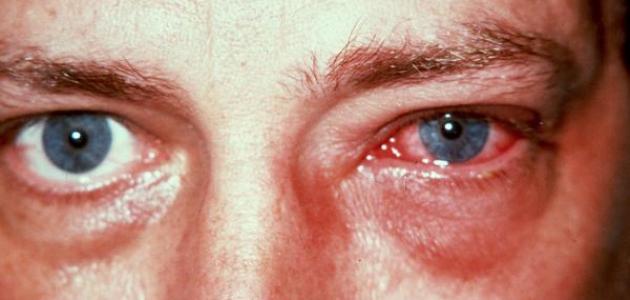
- Dry eye: occurs dry eye when there is a disturbance in the eye 's ability to produce tears naturally, as it leads to the difficulty of performing some daily activities, such as: reading, or use the computer for long periods.
References
- ↑ Liz Segre and Stephen Bagi, "Eye anatomy: A closer look at the parts of the eye" , www.allaboutvision.com , Retrieved 14-5-2019. Edited.
- ^ A b "Eye 's Anatomy , And Function" , Www.uofmhealth.org , 17-7-2018, Retrieved 14-5-2019. Edited.
- ↑ "Sclera" , www.healthline.com , 9-2-2015, Retrieved 14-5-2019. Edited.
- ↑ "How Does The Human Eye Work?" , www.nkcf.org , Retrieved 14-5-2019. Edited.
- ↑ Troy Bedinghaus, OD (1-6-2017), "What is the Choroid in Eye Anatomy?" , Www.verywellhealth.com , Retrieved 15-5-2019. Edited.
- ^ A b "How To Your Eye Works" , Lookafteryoureyes.org , Retrieved 15-5-2019. Edited.
- ↑ Ker Than (5-5-2016), "How the Human Eye Works" , www.livescience.com , Retrieved 15-5-2019. Edited.
- ↑ James Garrity (1-3-2019), "Protective Features of the Eyes" , www.msdmanuals.com , Retrieved 15-5-2019. Edited.
- ↑ "Age-Related Eye Diseases" , nei.nih.gov , Retrieved 15-5-2019. Edited.